2018年第45卷第8期目录
| |
|
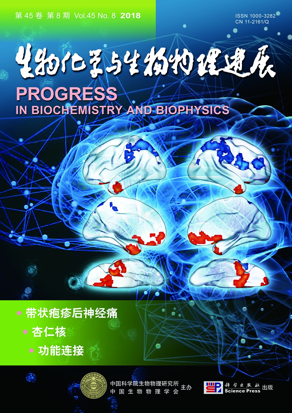
封面故事:带状疱疹后神经痛是一种常见的神经病理性疼痛,但其中枢机制尚不明了.杏仁核在疼痛反应中的作用近年来受到关注.以往研究尚未有将带状疱疹后神经痛患者杏仁核各个亚区进行功能连接分析,付维亮等通过功能磁共振成像研究带状疱疹后神经痛患者杏仁核各个亚区的功能连接的改变,探索慢性神经病理性疼痛的中枢机制.研究结果发现带状疱疹后神经痛患者杏仁核功能连接相较健康志愿者发生了改变,并与病程长短以及疼痛模拟评分表现出一定关联性,提示在慢性神经病理性疼痛的产生和发展中杏仁核以及多个涉及情绪、认知、注意的脑区发挥了重要作用,对于揭示和理解慢性神经病理性疼痛的中枢机制具有一定意义.
(付维亮,陶 蔚,陈富勇.带状疱疹后神经痛患者杏仁核的功能连接改变研究,本期第841~852页)
Cover Story:Postherpetic neuralgia (PHN) is a common type of neuropathic pain, the central mechanism of which is still unclear. The amygdala has recently garnered increased attention in pain processing. The purpose of this study is to investigate the functional neural networks of the amygdala in PHN and explore the mechanism of chronic neuropathic pain. Conventional magnetic resonance imaging (MRI) and resting-state functional MRI (fMRI) were performed in eight PHN patients and eight healthy controls. The functional connectivity (FC) of each subregion of the amygdala with the whole brain was computed. Paired t tests of the FC data were performed between the two experimental groups. Correlation analysis was applied between disease duration, visual analog scale (VAS), and FC strength. We found increased FC between the laterobasal (LB) and superficial (SF) amygdala and several brain regions including the temporal lobe and frontal lobe. We observed decreased FC between the SF amygdala and the precentral cortex, as well as the SF amygdala and parietal lobe. Correlation analysis showed that FC strength of the LB amygdala with both the temporal lobe and frontal lobe changed with disease duration and VAS in PHN patients. This altered FC in PHN suggests that the amygdala and several other brain regions involved in emotion, recognition, and attention play an important role in the modulation of chronic neuropathic pain.
|
综述与专论
研究报告
|
|
