|
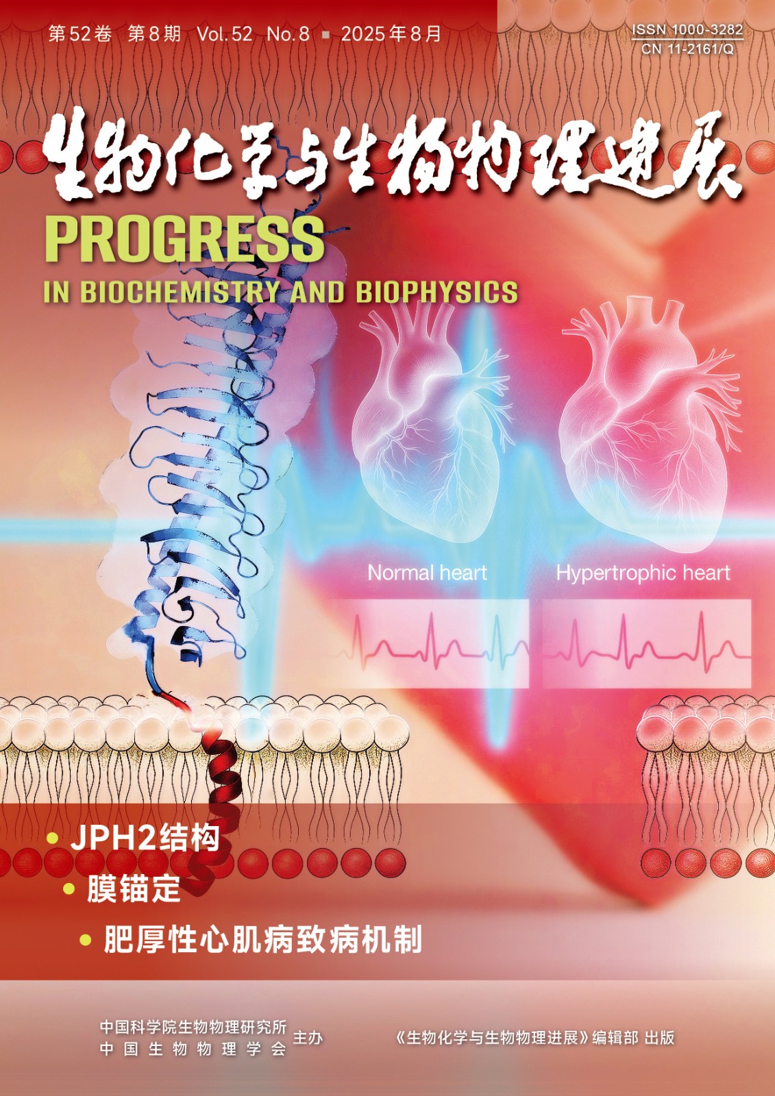
封面故事:该研究聚焦于心肌功能关键蛋白Junctophilin-2 (JPH2),旨在阐明其膜锚定机制及其在
肥厚型心肌病(HCM) 中的作用。JPH2是维持心肌细胞信号平台稳定的核心蛋白,但其N端如何
精确结合质膜尚不明确。研究团队通过X射线晶体学解析了JPH2 N端的高分辨率结构,发现其膜结
合的关键并非此前推测的MORN重复序列,而是一段富含正电荷的柔性连接区,该区域通过静电
作用将蛋白质锚定于膜表面。更重要的是,研究发现,HCM的一个关键致病突变位于此连接区,
该突变显著削弱JPH2的膜结合能力,导致其功能异常。这项研究不仅首次提供了JPH2的原子级结
构,还清晰构建了从“基因突变”到“蛋白质功能缺陷”再到“心肌疾病”的分子机制,为JPH2
相关心肌病的精准诊断和靶向治疗奠定了理论基础。
(王静欣,李志炜,刘伟,张文清,李健潮. Juntophilin-2 膜结合及肥厚型心肌病相关突变的结构基础,
本期第2103~2116 页)
Cover Story:Objective Junctophilin-2 (JPH2) is an essential structural protein that maintains junctional membrane complexes (JMCs) in cardiomyocytes by tethering the plasma membrane to the sarcoplasmic reticulum, thereby facilitating excitation-contraction (E-C) coupling. Mutations in JPH2 have been associated with hypertrophic cardiomyopathy (HCM), but the molecular mechanisms governing its membrane-binding properties and the functional relevance of its membrane occupation and recognition nexus (MORN) repeat motifs remain incompletely understood. This study aimed to elucidate the structural basis of JPH2 membrane association and its implications for HCM pathogenesis.Methods A recombinant N-terminal fragment of mouse JPH2 (residues 1-440), encompassing the MORN repeats and an adjacent helical region, was purified under near-physiological buffer conditions. X-ray crystallography was employed to determine the structure of the JPH2 MORN-Helix domain. Sequence conservation analysis across species and junctophilin isoforms was performed to assess the evolutionary conservation of key structural features. Functional membrane-binding assays were conducted using liposome co-sedimentation and cell-based localization studies in COS7 and HeLa cells. In addition, site-directed mutagenesis targeting positively charged residues and known HCM-associated mutations, including R347C, was used to evaluate their effects on membrane interaction and subcellular localization.Results The crystal structure of the mouse JPH2 MORN-Helix domain was resolved at 2.6 ?, revealing a compact, elongated architecture consisting of multiple tandem MORN motifs arranged in a curved configuration, forming a continuous hydrophobic core stabilized by alternating aromatic residues. A C-terminal α-helix further reinforced structural integrity. Conservation analysis identified the inner groove of the MORN array as a highly conserved surface, suggesting its role as a protein-binding interface. A flexible linker segment enriched in positively charged residues, located adjacent to the MORN motifs, was found to mediate direct electrostatic interactions with negatively charged phospholipid membranes. Functional assays demonstrated that mutation of these basic residues impaired membrane association, while the HCM-linked R347C mutation completely abolished membrane localization in cellular assays, despite preserving the overall MORN-Helix fold in structural modeling.Conclusion This study provides structural insight into the membrane-binding mechanism of the cardiomyocyte-specific protein JPH2, highlighting the dual roles of its MORN-Helix domain in membrane anchoring and protein interactions. The findings clarify the structural basis for membrane targeting via a positively charged linker and demonstrate that disruption of this interaction—such as that caused by the R347C mutation—likely contributes to HCM pathogenesis. These results not only enhance current understanding of JPH2 function in cardiac E-C coupling but also offer a structural framework for future investigations into the assembly and regulation of JMCs in both physiological and disease contexts.
|
